
There is a widespread concern that pregnant women should refuse to undergo an ultrasound examination, as it may harm the unborn child. We decided to check whether this fear is supported by scientific data.
ABOUT doubts it is safe, or at least not sufficiently proven to be safe, to perform ultrasound during pregnancy write authors thematic sites and large Media. Critics called an ultrasound procedure with a voluntary paid mutation for the unborn child, the consequences of which will appear in 15–20 years. Some sources clarifythat the potential harm may be higher at the beginning of the term, others indicate, that during pregnancy you should not do an ultrasound more than 12 times. They also write about the dangers of such diagnostics social network users and visitors thematic forums. And on sites with petitions, even require ban ultrasound for pregnant women (spelling and punctuation preserved): “Parents, fathers and mothers, think about it!!! Perhaps you have already seen the collection of money for expensive operations for children. Think about the suffering of so many families! All of them are caused by direct exposure to ultrasound.”
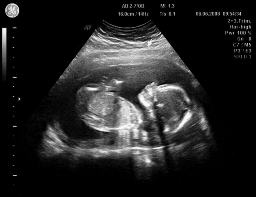
Ultrasound (US) is a non-invasive and widely available diagnostic method. During pregnancy it perform with several goals:
- confirm the presence of pregnancy, determine the number of embryos and the site of attachment of the fertilized egg, estimate the gestational age or identify complications such as ectopic pregnancy at an early stage;
- assess the dynamics of fetal development, clarify its heart rhythm and features of organ development;
- diagnose potential fetal malformations, monitor the condition of the placenta and umbilical cord;
- to clarify the development of fetuses in the case of multiple pregnancies, to assess the risk of premature birth.
According to modern WHO protocols, a woman recommended Carry out at least one ultrasound examination up to 24 weeks. Ministry of Health of the Russian Federation provides as many as three such screenings: at 11–13, 18–21 and 30–34 weeks.
The principle of operation of ultrasound is based on the emission of waves in range from 1 to 10 MHz. Waves are being sent to the area of interest, pass through the skin and other soft tissues, and are then reflected from internal organs and structures such as the fetus, placenta and amniotic fluid. Depending on its density and composition, each such structure will reflect waves differently. An ultrasound scanner receives reflected waves and converts them into an image on a monitor screen.
Many reputable medical organizations declare the safety of ultrasound for the body of a pregnant woman and the fetus - National Health Service UK, Centers for Disease Control and Prevention USA, Johns Hopkins University, clinics Cleveland And Mayo, American College of Obstetricians and Gynecologists. They all point out that ultrasonic waves, unlike X-ray diagnostics, do not provide radiation exposure to the body. Moreover, they cannot accumulate in the body, so ultrasound diagnostics can be carried out quite often.
The power of the device used during the procedure is extremely low. Candidate of Medical Sciences, gynecologist Vladimir Sursyakov leads the following data: modern devices operate in the range from 15 to 730 mW/cm²; for obstetric studies, a power of 180 mW/cm² is usually used. For comparison, power smartphone - about 200 mW, and Wi-Fi points - 100 mW. These devices operate in a constant emission and reception mode - they simultaneously receive and send waves. In contrast, an ultrasound machine emits waves only 20% of the time, and receives a signal 80% of the time. Thus, during ultrasound screening, a pregnant woman receives significantly less radiation than, for example, when watching a video or reading posts on social networks.
There is also no need to be afraid of tissue heating due to the effects of the device. Let’s say it operates at a power of 180 mW/cm2, under ideal conditions (that is, all the energy is converted into heat that enters the pregnant woman’s body, and is not dissipated in space). If the diagnostic duration is 30 minutes, then the device will spend only 6 minutes in radiation mode. (360 sec.). Thus, it will take 64.8 J, or approximately 15.5 calories, to heat the uterus, amniotic fluid and fetus. This amount of energy is enough to heat 15.5 ml of water by 1 °C. At the 12th week of pregnancy, the volume of only amniotic fluid amounts to 50 ml, and in addition to them, between the device and the fetus there is also the uterine lining itself, soft tissues and skin of the mother’s body. It turns out that the radiation power is not enough to heat the fruit even by tenths of a degree.
Some opponents of ultrasound diagnostics refer to a 2006 study conducted at Yale University. Authors of this work studied migration of neurons in the mouse embryonic brain under the influence of prenatal ultrasound exposure. Scientists found that in groups where fetuses were exposed to this effect, some neurons migrated from the depths to more superficial layers of the brain. Similar processes in humans, the researchers suggested, could lead to low birth weight, delayed speech development, and behavioral disorders.
Jacques Abramovich, doctor medicine and member of the World Federation of Ultrasound in Medicine and Biology, as well as the WHO task force on guidelines on non-ionizing radiation, criticized this is research. First, the mice were exposed to ultrasound radiation at 16.5–19.5 days of pregnancy (the entire pregnancy of mice lasts on average 19 days), that is, for women this would be 36–40 weeks, when such screenings are usually no longer carried out. Secondly, the mice were irradiated using not an obstetric ultrasound machine, but a device that is used in cardiology. Its power and range of operation are different, so it is not entirely correct to transfer the results of the study to the diagnosis of a pregnant woman. Finally, the volume of the mouse brain is much smaller than that of a human fetus at recommended diagnostic times. Moreover, in rodents the brain develops within a couple of days, while in humans this process lasts throughout pregnancy and after birth. So, half an hour of ultrasound influence on the brain of a human fetus in relation to the entire development time of the organ is significantly less than a similar influence on the mouse brain, and will not lead to similar consequences.
Another source argumentation, popular among opponents of ultrasound, is the book “Wave Genome” (1994). Its author, Pyotr Garyaev, claims that DNA is not a sequence of nucleotides, but a kind of wave, therefore, sound waves allegedly cause the genome to mutate. However, this concept fully pseudoscientific and is not based on any facts or research. Also Garyaev offers treat mutated cells with affirmations and supports the concept of telegony, according to which a child receives genetic information not only from his father, but also from all previous partners of the mother (“Verified” already told, why this theory has no basis).
At the same time, scientific data demonstrate the safety of ultrasound screening for the health of a pregnant woman and her unborn child. In 2011, the National Institute for Cancer Research (UK) analyzed 2690 cases of cancer in children under five years of age and their relationship with x-ray and ultrasound examinations performed both before birth and in the first 100 days of life. Exposure to X-rays modestly increased the risk of all cancers, especially leukemia, but there was no association between prenatal screening or ultrasound in the early days of life and an increased risk of cancer. Studying 512 cases of malignant brain tumors among children under 15 years of age, scientists from Uppsala University (Sweden) also not identified there is no pattern between the occurrence of the disease and the passage of ultrasound diagnostics, including while children are in the womb.
In 2017, WHO specialists summarized results of various studies on ultrasound diagnostics. After analyzing the outcomes of 35,737 births, experts noted that ultrasound before 24 weeks did not increase the risks of perinatal mortality and low birth weight. Late screening (30–36 weeks) does not affect this either, as data from another 30,675 births show.
Results population research indicate that children whose mothers had ultrasound scans during pregnancy are no different from other peers in physical development, school performance and the age of learning to read and write. The only identified difference — there are slightly more left-handers among them, especially among boys whose mothers were examined at 19–22 weeks. Scientists cannot yet explain whether ultrasound screening can influence the development of a dominant hand, and if so, why.
Thus, there is no scientific evidence that ultrasound examinations performed during pregnancy expose the fetus to radiation or otherwise adversely affect it. Children born after such screening are in no way inferior to their peers in physical and mental development. Ultrasound exposure in the prenatal period does not increase the risk of malignant tumors and leukemia. At the same time, ultrasound allows you to timely identify complications during pregnancy and thereby help both the pregnant woman and the unborn child.
Cover image: Image by Julio Cesar costa The Megament from Pixabay

If you find a spelling or grammatical error, please let us know by highlighting the error text and clicking Ctrl+Enter.