Many people believe that some medical research is harmful to health and can themselves cause various diseases. We decided to check whether these fears were justified.
Users scheduled for an MRI scan ask, is it dangerous, on the sites services questions and answers. On forums people share stories that they were not recommended to undergo an MRI due to possible harm. More like this questions calls the procedure radiography - Internet users often are interestedhow harmful it is and how often you can undergo it, share their fears about research. As for computed tomography, it scare even doctors. For example, Alexander Myasnikov statedthat one in 500 children undergoing this procedure will develop a brain tumor.
Magnetic resonance imaging (MRI)
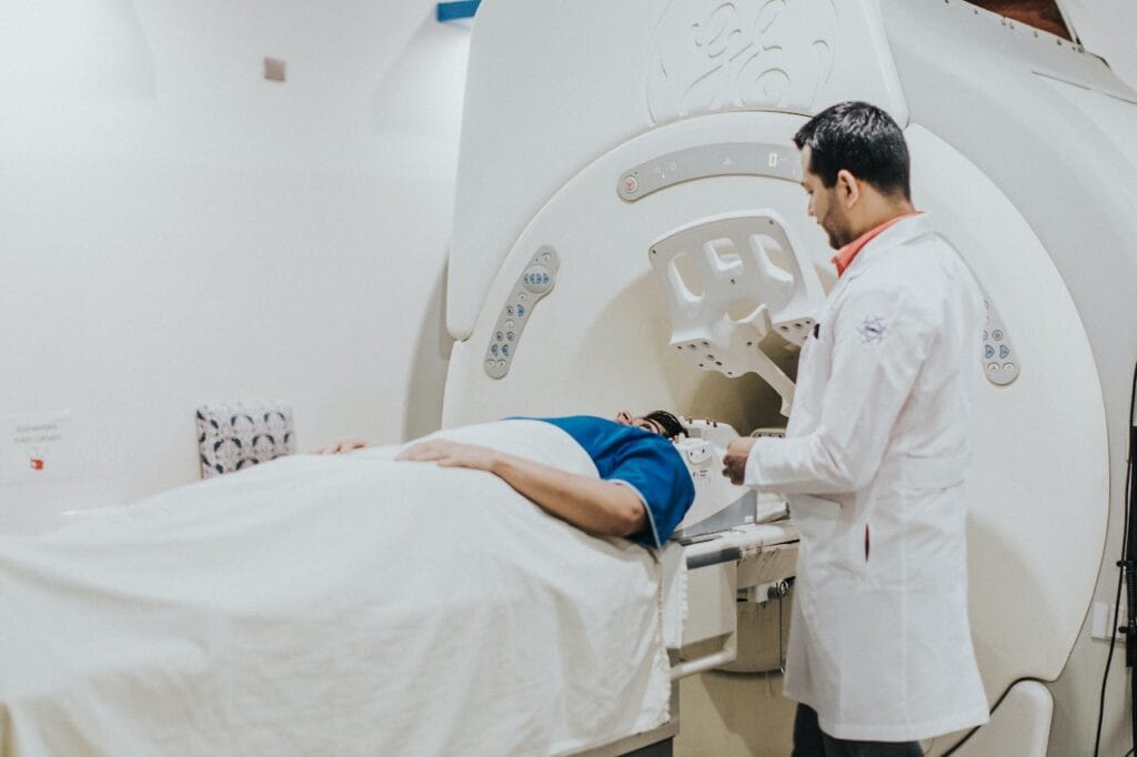
MRI Today it is one of the most effective methods of medical research. An MRI machine is a large tube with powerful magnets, under the influence of which protons in the patient’s body are arranged in a certain order. Short bursts of radio waves are then sent to the areas of the body being examined, knocking the protons out of balance. When the radio waves are turned off, the protons rearrange themselves again. Special receivers detect their movement - this helps to obtain information about the exact location of protons in the body and, as a result, an accurate picture of the structure of human internal organs. Protons in different types of tissue rearrange at different rates and produce different signals, so receivers are able to distinguish, for example, bones from muscles. The MRI procedure, unlike X-rays, does not use radiation, so from this point of view the procedure is absolutely safe.
Since the study involves powerful magnets, it is necessary to remove all metal objects. If the patient has any metal-containing implants (such as pacemakers, some types of prosthetics, pins or plates) or other objects, such as bullets or shrapnel, that cannot be removed, the MRI procedure may cause serious injury. This is one of the contraindications to conducting the study.
In some cases, to obtain more accurate information during MRI, the patient is given intravenous so-called contrast agent, which may cause an allergic reaction. It is also not recommended for use in severe renal impairment, since gadolinium, contained in some contrast agents, can provoke the development of nephrogenic systemic fibrosis, a disease that leads to damage to internal organs. However, experts note that all of these side effects from the use of contrast agents are very unlikely.
The use of contrast agents is not recommended and during pregnancy, since gadolinium increases risk of infant mortality and fetal development pathologies. As for MRI without contrast, there is no confirmed data on the negative impact of this procedure on a pregnant woman and child. However this question has not yet been well studied, and more research is needed to accurately answer the question of how safe it is.
In general, reputable medical organizations such as National Health Service UK, Stanford University (USA) and National Health Advisory Service In Australia, MRI is considered a completely safe procedure.
Thus, the MRI procedure has several contraindications. The main one is the presence of metal objects in the body that cannot be removed (for example, stuck fragments or a pacemaker). Using MRI, in these cases, you can get serious injuries, since the powerful magnets in the machine will attract all the metal to themselves. Contrast agents, sometimes used to improve diagnostic accuracy, can cause an allergic reaction. They also should not be administered to patients with severe renal impairment and should be used with caution during pregnancy. Moreover, in the absence of contraindications, experts call MRI a completely safe procedure that does not have any irreversible effects on human health.

Mostly not true
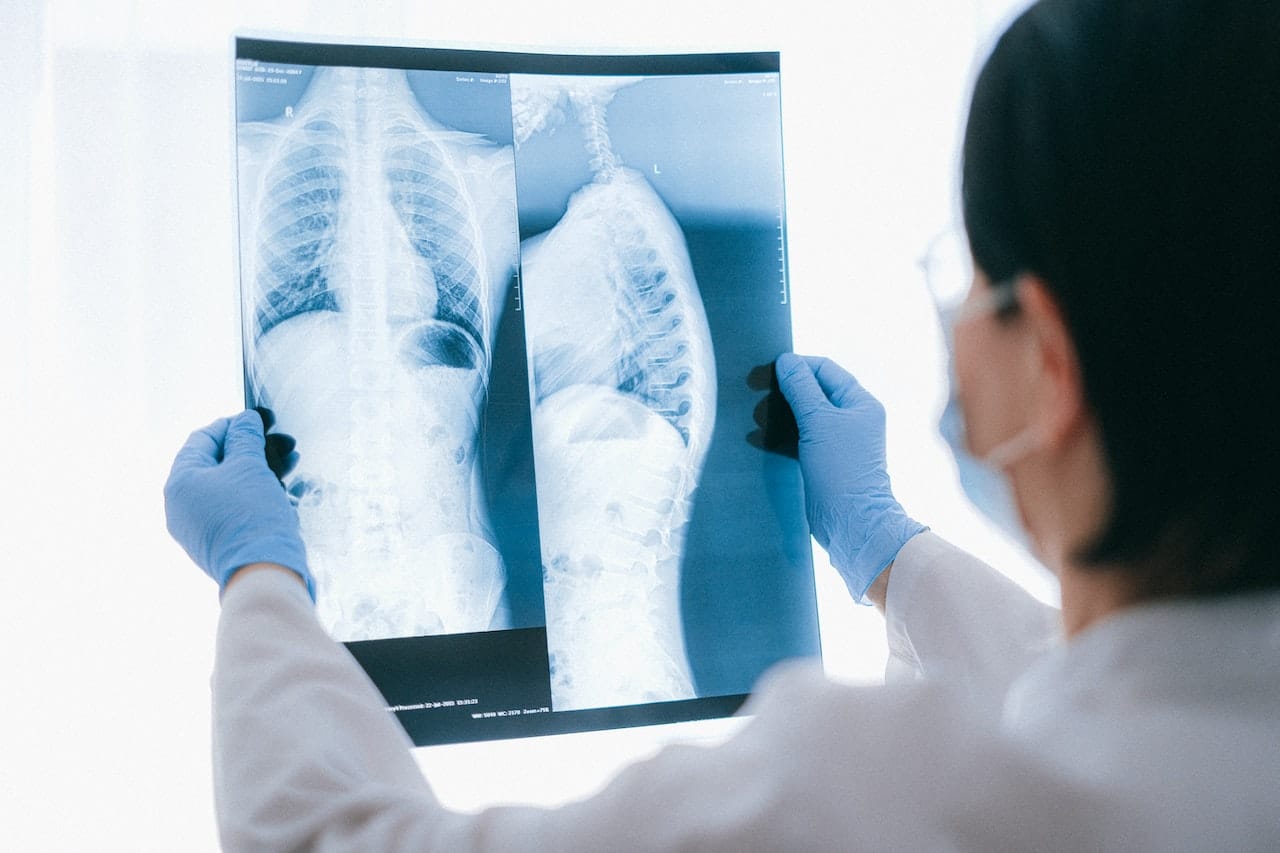
X-ray
X-ray allows you to obtain a black and white image of the internal structure of a person due to the influence of electromagnetic radiation. Different tissues absorb different amounts of radiation, and this affects the image on the image: the bones that absorb the most appear white, fat and muscle appear gray, and the lungs appear black.
When people hear the word “radiation,” they usually get scared, remembering the Chernobyl accident or the bombing of Hiroshima and Nagasaki, but natural background radiation is constantly present in the environment and it is impossible to avoid its exposure. For example, in USA On average, a person receives about 6.2 mSv from natural sources. In Russia in 2020 this indicator was about 4.18 mSv. If we compare the effects of x-rays with this background, then a chest examination comparable according to the received radiation dose with natural exposure for approximately ten days. And this dose is still three times lower than what get passengers of long-haul flights.
The radiation dose also depends on what part of the body is being examined. For example, specialists from the American Dental Association claimthat by taking an X-ray of the jaw, patients receive significantly less radiation compared to daily natural exposure.
At the same time, too frequent X-ray examinations can actually be harmful to health. That is why doctors recommended record such studies in patients’ medical records in order to compare the need for x-rays and the possible harm from it. Experts note that x-rays can indeed provoke the development of cancer in the long term, but this risk extremely small and again depends on the area of study. For example, an x-ray of an arm or leg can cause cancer in only one person out of a million, but the lumbar spine can cause cancer in one out of 15,000. barium - a contrast agent that allows X-ray examination of, for example, the abdominal cavity - seriously increases these risks. However, proven promotion The risk of developing cancer occurs only when receiving a dose of 100 mSv per year.
Thus, if X-rays are taken only when necessary - for example, for fractures or dental treatment - the health risk is minimal. Potential harm depends on the area of study, some of which are comparable in exposure to natural background radiation that people receive daily from the environment. In addition, doctors typically use lead aprons when conducting this type of research to protect other parts of the body from radiation, which also reduces exposure. However, some types of exposure, especially those involving barium-based solutions, may actually increase the likelihood of developing cancer, albeit slightly.

Half-truth
Computed tomography (CT)

CT - this is a more advanced type of x-ray examination using many rays that pass through the organ being examined at different angles and allow you to obtain a more detailed image. Accordingly, all the risks of X-rays associated with radiation are also true for CT. Moreover, the dose of radiation that the patient receives during such an examination is significantly higher than with a conventional x-ray. For example, during a CT scan of the abdomen, a person receives an exposure equal to four and a half years of life in a natural radiation background (with ordinary X-rays - about four months). Accordingly, the risk of developing cancer due to such a study is higher: it can develop in one person out of 2000.
Thus, unlike X-rays, the long-term risks of developing cancer from CT scans are significantly higher. However, diagnostics using this technology are more accurate. Therefore, doctors in each specific case decide on the advisability of conducting a particular study, weighing the risks and potential benefits.
Half-truth
Fluorography
Fluorography - another type of x-ray examination, used, in particular, to detect tuberculosis and other pulmonary diseases.
In Russia, fluorography is included in the protocol medical examinationwhich doctors advise to undergo regularly. However, WHO back in 1974 called abandon mass regular x-ray examinations due to the fact that the percentage of patients with tuberculosis or other diseases identified using this method is too small.
At the same time dose The radiation received during the chest fluorography procedure is small - only about 0.5 mSv for film shooting and 0.05 mSv for digital shooting. This is comparable in terms of the impact on the body with conventional x-rays, which was already mentioned above. Although theoretically fluorography, like other studies using radiation, can increase the risk of developing cancer, experts do not consider this danger to be significant.
Thus, although fluorography, like X-rays, exposes the human body to radiation and this is not beneficial, it has a negligible impact on health. Moreover, in some countries, including Russia, fluorography is included in the clinical examination protocol and is recommended to be performed regularly. However, WHO experts recommend abandoning this practice, because the benefits do not exceed the theoretical harm and costs of conducting such studies.
Half-truth
Cover photo: Anna Shvets, pexels.com
Read on the topic:
- Is it true that anesthesia takes away five years of your life?
- Is it true that COVID-19 vaccination has led to a sharp increase in the incidence of myocarditis?
- Is it true that you need to take iodine immediately after exposure to radiation to protect against infection?
If you find a spelling or grammatical error, please let us know by highlighting the error text and clicking Ctrl+Enter.