
In media publications and bloggers you can find an amazing story: a Frenchman lived for many years with only 10% of his brain, while feeling well, and the pathology was discovered only when he complained of weakness in his legs. We decided to check if it was true.
Many media outlets wrote about the Frenchman with 10% of the brain: “Komsomolskaya Pravda"(A Frenchman lives for 50 years without a brain"), "Moscow 24"(A Frenchman has lived without a brain for 50 years"), "News"("A Frenchman lived for 44 years with virtually no brain") or Lenta.ru (“The 44-year-old Frenchman has almost lost his brain”). This story was also mentioned in their materials Republic And "Russian newspaper" Briefly they tell like this: “A man is missing 90% of his brain. He collapsed within 30 years, but that didn’t stop him from working, getting married and having children.”
Without a detailed understanding of how the human brain works, it will not be possible to understand history. To make it easier to understand, structures in this explanation that are directly related to the story will be italicized.
The brain is an organ of the central nervous system located in the anterior and upper part of the cranial cavity. Brain of the average adult person weighs approximately 1200–1400 g and occupies the vast majority of the skull’s capacity.
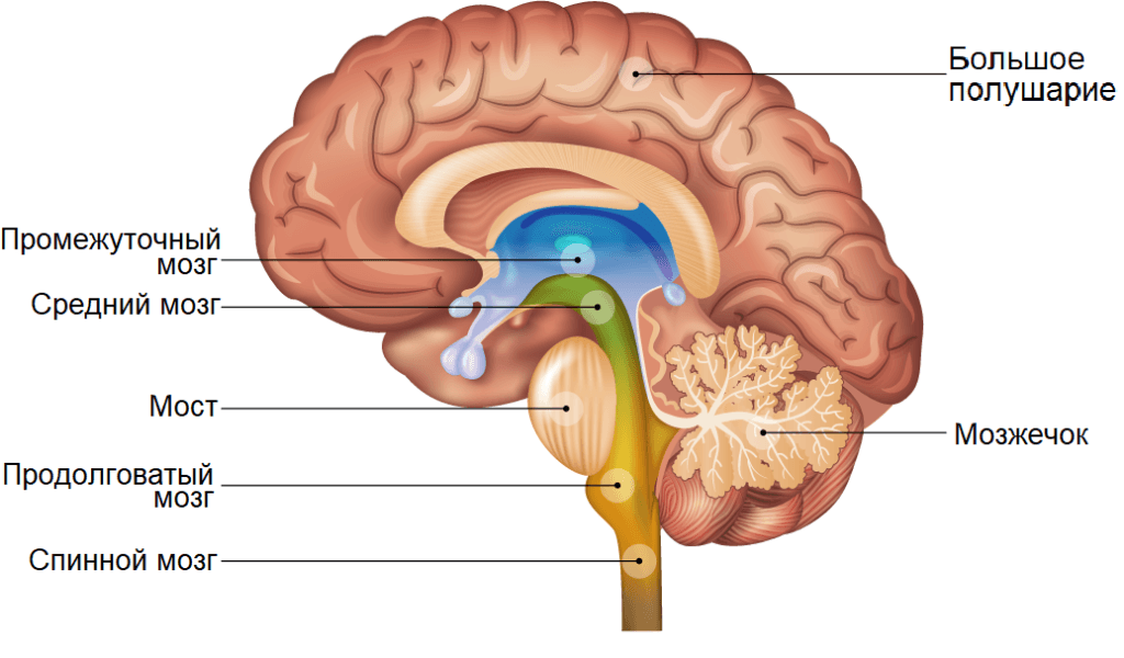
Accepted divide the brain into three large parts: cerebral hemispheres, cerebellum (Latin cerebellum - literally “small brain”) and brain stem. Also exists division into five departments:
- medulla oblongata;
- posterior, which includes the pons, cerebellum and pineal gland;
- average;
- intermediate;
- the forebrain, represented by the cerebral hemispheres.
In addition to the structures mentioned, in the brain also are ventricles are cavities filled with cerebrospinal fluid. There are four of them in total: two lateral ones (the left one is traditionally called the first, and the right - the second) communicate through the Monroe foramen with third, and that, in turn, through the Sylvian aqueduct with fourth. From the fourth ventricle, cerebrospinal fluid enters the subarachnoid space (the cavity between the pia mater and the arachnoid mater of the brain) through two lateral foramina of Luschka and one medially located Magendie hole (in the illustration this is the very bottom of the purple area).
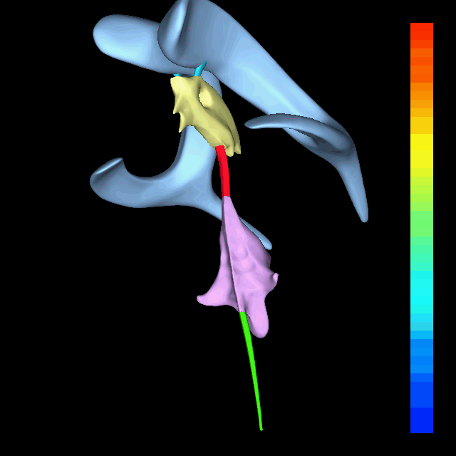
blue - lateral ventricles;
blue—Monroe foramen;
yellow - third ventricle;
red - Sylvian aqueduct;
purple - fourth ventricle;
green - transition to the central spinal canal.
en: Anatomography, CC BY-SA 2.1 JP, via Wikimedia Commons
Thus, the key concepts are the cerebrum, cerebellum and brainstem, as well as the third and fourth ventricles and the foramen of Magendie.
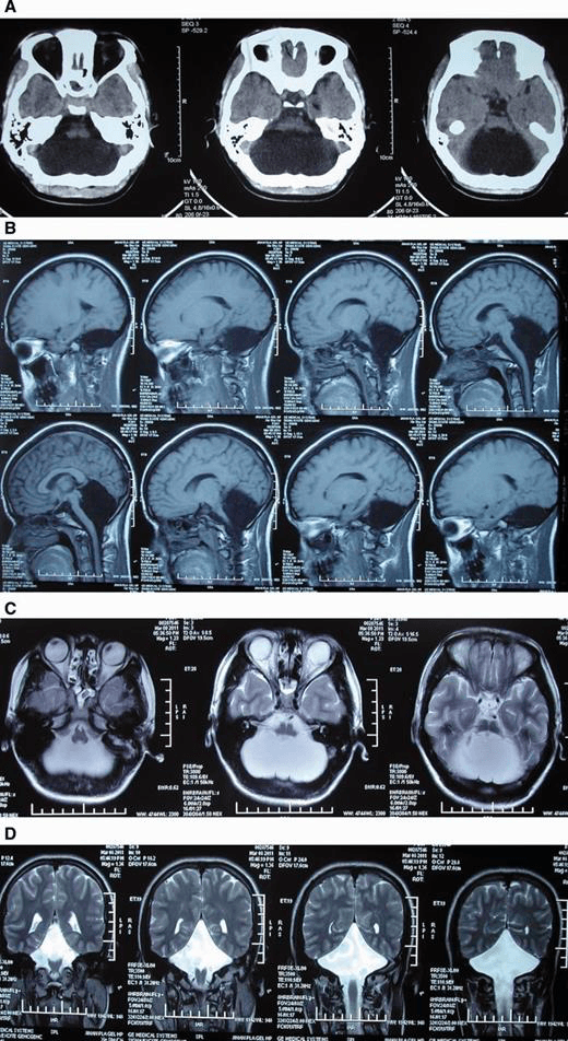
Some resources, for example "Komsomolskaya Pravda", in their publications they provided links to the original publication in the authoritative British medical journal The Lancet. There the case is described in the format of a clinical picture, that is, one paragraph of text and an accompanying photograph. The text, although small, is difficult to understand for non-specialists, so we will retell it in our own words.
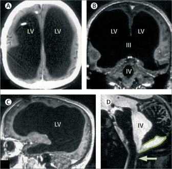
A 44-year-old man presented with a two-week history of mild weakness in his left leg. At the age of six months, the patient developed an accumulation of cerebrospinal fluid in the brain. Doctors installed a ventriculoperitoneal shunt - a tube through which excess fluid leaves the brain, is drained into the abdominal cavity and is absorbed into the venous bed. However, at the age of 14, the patient complained of poor coordination of movements and muscle weakness, so the shunt was removed (usually the shunt is installed permanently, but in some cases doctors may decide it is no longer needed and remove it. It is most likely that the child was closely monitored after the shunt was removed, but there was no evidence of cerebrospinal fluid accumulation. — Approx. ed.). His neurological development and medical history were normal. He is married, has two children and works as a government employee (in the original white collar, that is, “white collar”). Further, due to treatment as an adult, the patient underwent CT and MRI, where they discovered an enlargement of the lateral, third and fourth ventricles, a very thin cortical layer, the presence of a posterior fossa cyst and a narrowing of the foramen of Magendie. Neuropsychological testing showed that his intelligence quotient (IQ) was 75, with a verbal IQ of 84 and a performative IQ of 70. To help him, doctors performed a neuroendoscopic ventriculocisternostomy, that is, a minimally invasive hole was made in the third ventricle (yellow on the image) to prevent fluid from accumulating. However, the manipulation did not give the desired effect, and then he was again installed with a ventriculoperitoneal shunt, which dumps excess cerebrospinal fluid into the abdominal cavity. After insertion of the shunt, neurological examination results became normal within several weeks. The results of neuropsychological testing and CT scans were unchanged.
The case is indeed quite unusual, which is why it was published in such a reputable journal. However, scientists themselves do not write anything about 10% of the brain. In order to understand where this thesis comes from, remember that the brain is divided into three parts: the cerebrum, the cerebellum and the brainstem. Having studied the CT and MRI of the patient presented in the publication, the editor-in-chief of the neuronovosti.ru portal, scientific journalist and popularizer of science Alexey Paevsky notedthat the patient’s cerebellum and brain stem are completely intact and not changed in size. As a percentage of the weight of the entire brain, the cerebellum and brainstem make up about 13%, plus some part of the cerebrum (insignificant, but not zero) remained in the patient. That is, 10% is still an understatement; most likely, he retained at least 15% of the average brain weight.
The second important aspect that most publications miss is that neurons in the human brain are distributed extremely unevenly. It is in the cerebellum that they are most numerous. By estimates According to some scientists, of the 120 billion neurons in the brain, 101 billion are located in the cerebellum. Other evaluate the total number of neurons is more modest: “only” 85 billion, of which 69 billion are again in the cerebellum. Thus, if we translate brain loss from weight and volume into the number of neurons, the figure will not be so shocking - the Frenchman lost approximately 20% of all neurons.
Unfortunately, in The Lancet, scientists do not say at what age the patient’s brain began to be replaced by cerebrospinal fluid. It is most likely that before the discovery of this pathology, neither CT nor MRI diagnostics were performed on him, so there is no way to know what his brain looked like at six months or at 14 years. Moreover, there is not enough data to even understand whether he was born with this feature or whether the cerebrospinal fluid gradually dissolved his brain.
It is also important to keep in mind the so-called neuroplasticity - with the congenital absence or loss of part of the brain and neurons, the remaining areas are able to fully or partially take over its functions. Here are a couple of examples.
A 24-year-old Chinese woman consulted doctors about persistent dizziness, and specialists found outthat she has been deprived of the cerebellum since birth. Instead, the woman's brain contained only cerebrospinal fluid. Moreover, at that time she was already married and raising a daughter. It was noted that as a child she lagged behind her peers in development - she learned to stand only at four years (on average, this happens at nine to ten months), and to walk without support at seven years (on average, this happens at about 12 months). However, as an adult, the woman almost ceased to differ from her peers. She doesn't speak clearly and can't always touch the tip of her nose with her eyes closed, but despite missing 80% of her neurons, she lives a completely normal life.
People can live completely without one hemisphere of the brain, surgically removed. Such an operation called hemispherectomy, and most often it carry out, to rid patient from drug-intractable seizures epilepsy. American scientists compared MRI of the brains of six adults who had undergone such surgery as children, with six volunteers. It turned out that in patients after hemispherectomy, connections in the brain do not differ significantly from normal, and cognitive and physical abilities are the same as those of their peers. Loss of one hemisphere at an early age stimulates development of the second, and the person does not experience any special problems. The operation of hemispherectomy in adulthood and its consequences is more or less plausible shown in the 15th episode of the third season of the popular television series House.
Thus, in most details the story about the Frenchman who lost a significant part of his brain is correct. However, journalists very freely calculated this 10%: 13%, accounting for the cerebellum and brain stem, are completely intact, and the patient also has a small part of the cerebral cortex left. Moreover, in terms of the number of lost neurons, the situation is even less fatal - 80% of the neurons are in the completely preserved cerebellum, that is, the loss was less than 20%. The most accurate (though not as impressive) headline for such news would be: “A Frenchman lived for decades with only 10% of his cerebral cortex.”
Cover image: Image by Pexels from Pixabay

Mostly true
If you find a spelling or grammatical error, please let us know by highlighting the error text and clicking Ctrl+Enter.